Home » Uncategories » Blood Vessels Labeled : 27 best Practical II images on Pinterest : Vessel networks deliver blood to all tissues in a directed and regulated manner.
Tuesday, 1 June 2021
Blood Vessels Labeled : 27 best Practical II images on Pinterest : Vessel networks deliver blood to all tissues in a directed and regulated manner.
Blood Vessels Labeled : 27 best Practical II images on Pinterest : Vessel networks deliver blood to all tissues in a directed and regulated manner.. The vessels that carry blood away from the heart are called arteries, and their very small branches are arterioles. Anatomy of blood vessels review sheet 32 261 microscopic structure of the blood vessels 1. Blood vessels form a continuous path for blood flow that starts and ends at the heart.arteries carry blood away from the heart, regardless of the degree of blood oxygenation.veins carry blood toward the heart. When sphincter muscles are relaxed, the capillary bed is open, and blood flows through the capillaries. Digestive system worksheet answers 12 photos of the digestive system worksheet answers digestive system worksheet answer key, digestive system worksheet pearson education, human digestive system worksheet with answers, the digestive system digestion and absorption worksheet answers, the human digestive.
The smallest veins are called venules. Blood vessel structure and function cardiovascular system: The adventitia or outer layer which provides structural support and shape to the vessel Related posts of the human blood vessels labeled digestive system worksheet answers. Start studying blood vessels labeling.
Circulatory Pathways · Anatomy and Physiology from philschatz.com Identify the structural layers of arteries and veins. Between arteries and veins, there is a network of. Blood vessels consist of arteries, arterioles, capillaries, venules, and veins. This article lists a series of labeled imaging anatomy cases by system and modality. Figure 12.11 anatomy of a capillary bed. Interactive physiology with quizzes cardiovascular system: The function and structure of each segment of the peripheral vascular system vary depending on the organ it supplies. Name the blood vessel labeled 'd'.
When sphincter muscles are relaxed, the capillary bed is open, and blood flows through the capillaries.
These processes can be primary or secondary, and they tend to affect only vessels of a specific type or in a specific location. Distinguish between the structure of arteries and veins. Name the blood vessel labeled 'd'. Free online quiz blood vessel labeling Vessel networks deliver blood to all tissues in a directed and regulated manner. Figure 12.11 anatomy of a capillary bed. Deoxygenated blood from the peripheral veins is transported back to the heart from capillaries, to venules, to veins, to the right side of the heart, and then. Arteries, arterioles, capillaries, venules and veins. There are five main types of blood vessels: Blood is supplied to parts within the neck, head and brain through branches of the subclavian and common carotid arteries. The iliac, femoral, popliteal and tibial (calf) veins are the deep veins in the legs. Blood vessels are found throughout the body. Its smooth surface decreases resistance to blood flow
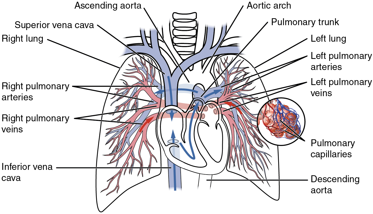
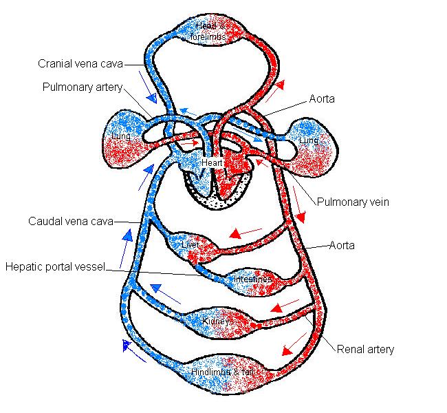
Figures 1 and 2 show the major arteries and veins of the body. Blood vessels consist of arteries, arterioles, capillaries, venules, and veins. Closed circulation of blood throughout the body deliver oxygen and nutrients to tissues. Aside from capillaries, blood vessels are all made of three layers: •formed where capillaries unite • extremely porous 1) venules:
Lab Exercise: Anatomy of Blood Vessels. Blood Vessel ... from reader012.fdocuments.in The major veins in the These processes can be primary or secondary, and they tend to affect only vessels of a specific type or in a specific location. There are five main types of blood vessels: Measuring blood pressure cardiovascular system: Very small branches that collect the blood from the various organs and parts are called venules, and they unite to form veins, which return the blood to the heart. This article lists a series of labeled imaging anatomy cases by system and modality. Veins (in blue) are the blood vessels that return blood to the heart. A primary purpose and significant role of the vasculature is its participation in oxygenating the body.
It extends on each side of the neck and divides at the level of the larynx into two branches:
A blood vessel's main function is to transport blood around the body. Start studying blood vessels labeling. Identify the structural layers of arteries and veins. The superior vena cava is not labeled in figure 7.4. Inner layer is made of simple squamous. Eventually, the smallest arteries, vessels called arterioles, further branch into tiny capillaries, where nutrients and wastes are exchanged, and then combine with other vessels that exit capillaries to form venules, small blood vessels that carry blood to a vein, a larger blood vessel that returns blood to the heart. Bios255 week 3 cardiovascular system: Arteries (in red) are the blood vessels that deliver blood to the body. This set is often in folders with. Closed circulation of blood throughout the body deliver oxygen and nutrients to tissues. When sphincter muscles are relaxed, the capillary bed is open, and blood flows through the capillaries. Aside from capillaries, blood vessels are all made of three layers: Blood vessel labeling online quiz;
Figures 1 and 2 show the major arteries and veins of the body. Vessel networks deliver blood to all tissues in a directed and regulated manner. Like arteries, veins form a complex, branching system of larger and smaller vessels. Name the blood vessel labeled 'd'. The inferior vena cava is labeled in the figure below.
The Anatomy and Physiology of Animals/Circulatory System ... from wikieducator.org The three major types of blood vessels: Measuring blood pressure cardiovascular system: The thick outermost layer of a vessel (tunica adventitia or tunica externa) is made of connective tissue. Name the blood vessel labeled 'c'. Between arteries and veins, there is a network of. 4 but is clearly visible entering the right atrium of the heart. Name the blood vessel labeled 'b'. Vessels transport nutrients to organs/tissues and to transport wastes away from organs/tissues in the blood.
Aside from capillaries, blood vessels are all made of three layers:
Name the blood vessel labeled 'b'. The iliac, femoral, popliteal and tibial (calf) veins are the deep veins in the legs. Best quiz blood vessel labeling; Arteries, veins, and capillaries blood vessels flow blood throughout the body. Figure 12.11 anatomy of a capillary bed. Inner layer is made of simple squamous. The 4 valves are the aortic, pulmonary, mitral, and tricuspid valves. Name the blood vessel labeled 'd'. Measuring blood pressure cardiovascular system: Blood vessels also play a role in controlling your blood pressure. Bulky middle tunic contains smooth muscle and elastin 3. The superior vena cava is not labeled in figure 7.4. Learn vocabulary, terms, and more with flashcards, games, and other study tools.
0 Response to "Blood Vessels Labeled : 27 best Practical II images on Pinterest : Vessel networks deliver blood to all tissues in a directed and regulated manner."


0 Response to "Blood Vessels Labeled : 27 best Practical II images on Pinterest : Vessel networks deliver blood to all tissues in a directed and regulated manner."
Post a Comment